Benefits of Cone Beam CT Scans
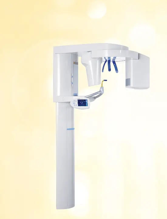
South Bay Dentistry harnesses the power of i-CAT® 3D Imaging to deliver exceptional dental care to our patients. Being a professional dental office in Gardena, we prioritize the integration of advanced technologies that enhance our diagnostic capabilities and treatment outcomes. i-CAT® 3D Imaging stands at the forefront of dental imaging technology, allowing us to capture detailed three-dimensional images of the oral and maxillofacial structures.
With this cutting-edge technology, we can accurately assess complex dental conditions, including impacted teeth, bone structure, and airway analysis. The high-resolution images generated by i-CAT® enable our skilled team of dental professionals to develop precise treatment plans, provide accurate diagnoses, and optimize the delivery of our comprehensive dental services. By incorporating i-CAT® 3D Imaging into our practice, we ensure that our patients receive the highest standard of care in a comfortable and efficient manner.
Oral surgery is a complicated field. One of the most important aspects is diagnosing the problem in a way that is accurate and effective.
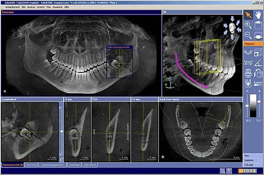
The utility of 3D imaging technology in dentistry is evident due to its unparalleled ability to unveil previously unseen details. Through this advanced technology, dental professionals gain access to cross-sectional views, including bucco-lingual perspectives, as well as axial, sagittal, coronal, and panoramic views. These comprehensive visualizations, made possible by digital technology, offer valuable insights that were previously unattainable. By utilizing 3D imaging, dental practitioners are equipped with a powerful tool to enhance diagnosis, treatment planning, and patient communication. This transformative technology empowers dental professionals to provide precise and effective care, revolutionizing the field of dentistry.
At South Bay Dental, our practice utilizes state-of-the-art 3D cone beam CT technology that provides highly accurate 3-D images. This technology can be used to diagnose dental implantology, TMJ analysis, airway assessment, and oral and orthognathic surgery.


i-CAT® 3D Imaging
- Dental implants
- Size and shape of ridge, quantity and quality of bone
- Number, orientation of implants
- Orthognathic surgery planning
- Oral and maxillofacial pathology
- Need for bone graft, sinus lift
- 3D CT ScanUse of implant planning software
- Oral and maxillofacial surgery
- Location of anatomic structures: mandibular canal, submandibular fossa, incisive canal, maxillary sinus
- Localization of impacted teeth, foreign objects, extra teeth
- Evaluation of facial fractures & asymmetry
- Localization and characterization of lesions in the jaws
- Relationship of lesion to teeth and other structures

